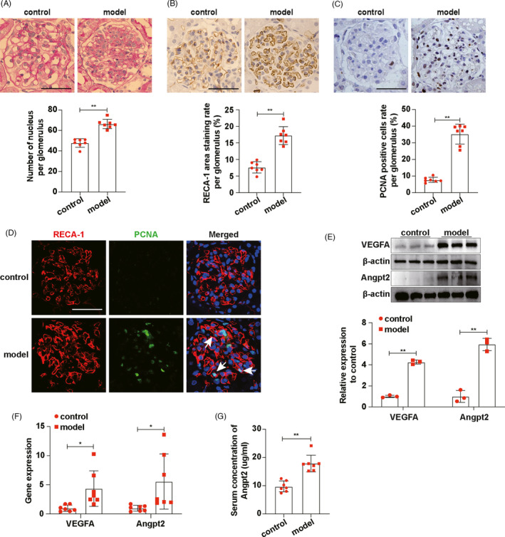
FIGURE 1.

On the 7th day of anti‐Thy‐1 nephritis, ECs proliferated and the expressions of VEGFA and Angpt2 increased. A, PAS staining showed glomerulopathy, and the number of nucleus per glomerulus was qualified (n = 7). B, RECA‐1 expressions in glomeruli were detected by immunohistochemical staining and RECA‐1 area staining rate per glomerulus was qualified (n = 7). C, PCNA expressions in glomeruli were detected by immunohistochemical staining and PCNA‐positive cells rate per glomerulus was qualified (n = 7). D, Locations of RECA‐1 and PCNA expressions were showed by immunofluorescence co‐staining. White arrows indicated proliferating ECs. RECA‐1, red. PCNA, green. DAPI, blue. E, VEGFA and Angpt2 expressions in glomeruli were detected by Western blot. F, mRNA expression levels of VEGFA and Angpt2 in glomeruli were detected by RT‐qPCR (n = 7). G, Concentrations of Angpt2 in rat serum were determined by ELISA (n = 7). Scale bars, 50 μm. Results are presented as the mean values (±SD), *P < .05, **P < .01. Angpt2, angiopoietin2; ECs, endothelial cells; PCNA, proliferative cells nuclear antigen; RECA‐1, rat endothelial cells antigen‐1; VEGFA, vascular endothelial growth factor A. Control, normal rats. Model, anti‐Thy‐1 nephritis rats
