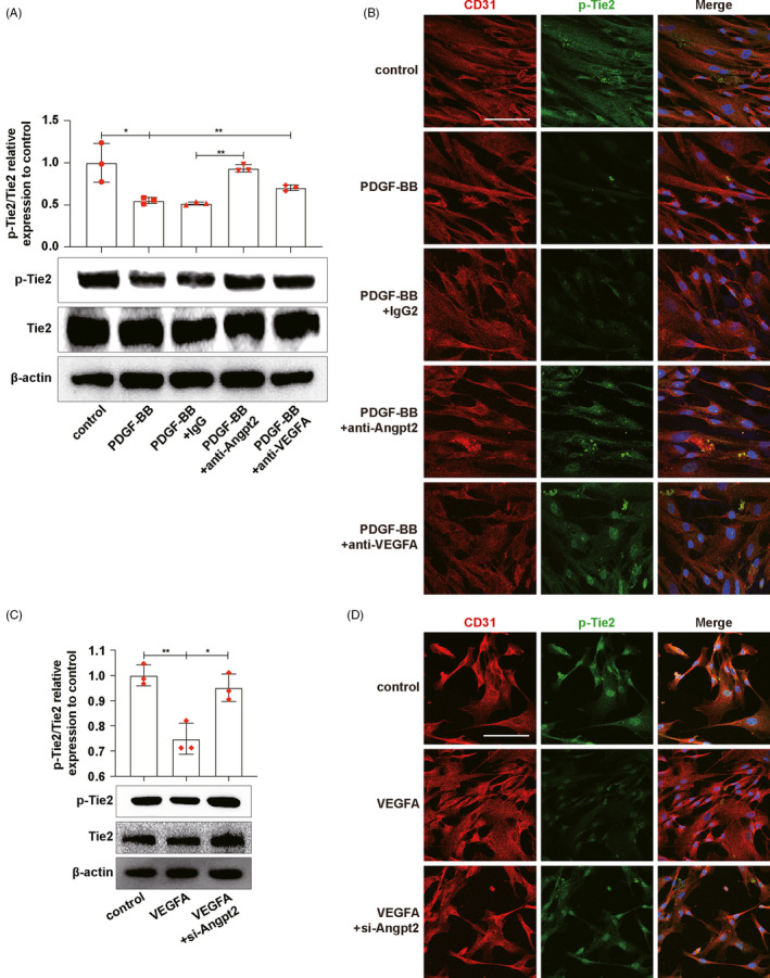
FIGURE 4.

MCs‐derived VEGFA stimulated Angpt2 expression which inhibited Tie2 phosphorylation on ECs. A, C, p‐Tie2 and Tie2 expressions on ECs were detected by Western blot. B, D, p‐Tie2 expressions on ECs were detected by p‐Tie2/CD31 immunofluorescence co‐staining. CD31, red. p‐Tie2, green. DAPI, blue. Results are presented as the mean values (±SD), *P < .05, **P < .01. Scale bars, 50 μm. Angpt2, angiopoietin2; ECs, endothelial cells; MCs, mesangial cells; PDGF‐BB, platelet‐derived growth factor BB, to activate MCs; Tie2, TEK tyrosine kinase. (A, B) Control, ECs co‐cultured with inactive MCs. PDGF‐BB, ECs co‐cultured with PDGF‐BB‐activated MCs. PDGF‐BB+IgG, ECs co‐cultured with PDGF‐BB‐activated MCs and IgG antibody, was added as negative control. PDGF‐BB+anti‐Angpt2, ECs co‐cultured with PDGF‐BB‐activated MCs and Angpt2 neutralizing antibody, was added. PDGF‐BB+anti‐VEGFA, ECs co‐cultured with PDGF‐BB‐activated MCs and VEGFA neutralizing antibody, was added. (C, D) Control, inactive ECs. VEGFA, ECs stimulated by VEGFA for 24 h. VEGFA+si‐Angpt2, ECs stimulated by VEGFA for 24 h after transfected with si‐Angpt2
