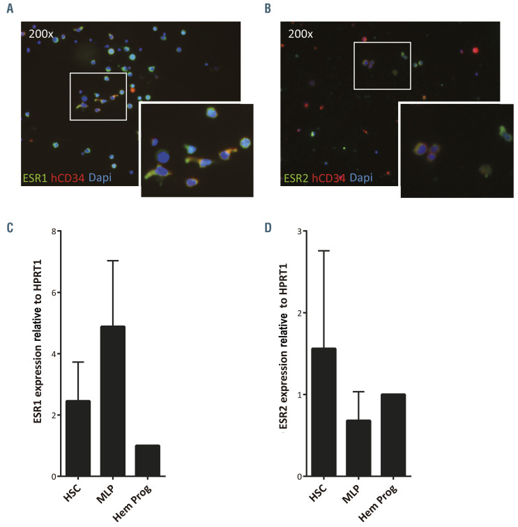
Figure 2.
Human hematopoietic stem and progenitor cells express estrogen receptors. (A) Representative immunofluorescent image of umbilical cord blood CD34+ (CB-CD34+) cells stained with anti-ESR1 (green), anti-hCD34 (red) and DAPI (blue). The insert, showing ESR1+ CD34+ cells (marked with arrows), is an enlargement of the white boxed area. (B) Representative immunofluorescent image of CB-CD34+ cells stained with anti-ESR2 (green), anti-hCD34 (red) and DAPI (blue). The insert, showing ESR2+ CD34+ cells (marked with arrows), is an enlargement of the white boxed area. (C) Qualitative real-time polymerase chain reaction (qRT-PCR) analysis of ESR1 expression of sorted hematopoietic stem cells/multipotent rogenitors (HSC/MPP: hCD34+hCD38-hCD45RA-), multilymphoid progenitors (MLP: hCD34+hCD38-hCD45RA+) and committed hematopoietic progenitors (Hem Prog: hCD34+hCD38+). (D) qRT-PCR analysis of ESR2 expression of sorted HSC/MPP, MLP and hematopoietic progenitors. Data were obtained from three biological replicates and are presented as the mean ± standard deviation. Statistical significance was analyzed by one-way analysis of variance with the Fisher least significant difference test: no significant differences were found.

