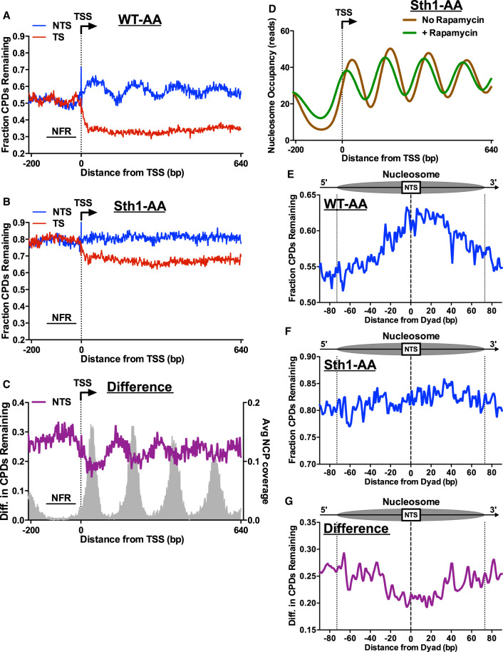
Figure 5.
RSC promotes repair of both linker and nucleosomal DNA. CPD-seq analysis examining the repair of CPDs on both the TS and NTS of approximately 5200 yeast genes in WT-AA (A) and Sth1-AA (B). Data plotted for each nucleotide position spanning −200 bp upstream of and +640 bp downstream from the TSS. The fraction of CPDs remaining is calculated as the ratio of damage after 2 h of repair compared with the damage immediately following UV irradiation (0 h). (C) Plot depicting the difference in unrepaired CPDs in Sth1-AA cells compared with WT-AA cells. Difference in unrepaired CPDs was smoothed using a 3-nt window. Gray peaks correspond to the sequencing coverage of dyad positions for position nucleosomes (Weiner et al. 2015). (D) Nucleosome occupancy plot of Sth1-AA cells with and without rapamycin treatment to visualize nucleosome shifting that is known to occur in RSC-depleted cells. Data taken from Kubik et al. (2018). (E,F) Analysis of CPD repair within nucleosomes. Repair of the NTS of WT-AA and Sth1-AA cells was examined for nucleosomes in yeast genes to examine potential GG-NER defects in RSC-depleted cells. Dotted lines at positions −73 and +73 bp from the dyad center indicate the boundary of the nucleosome core particle. Nucleosome positions for WT-AA are from vehicle-treated Sth1-AA cells, whereas those for Sth1-AA are from rapamycin-treated Sth1-AA cells (Kubik et al. 2018). (G) Plot depicting the difference in unrepaired CPDs within nucleosomes in Sth1-AA cells compared with WT-AA cells. Difference in unrepaired CPDs was smoothed using a 3-nt window.

