Figure 2:

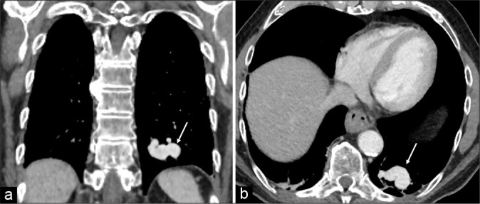
Coronal (a) and axial (b) chest computed tomography with intravenous contract shows large pulmonary arteriovenous malformation (arrow) in the left lower lobe.

Coronal (a) and axial (b) chest computed tomography with intravenous contract shows large pulmonary arteriovenous malformation (arrow) in the left lower lobe.