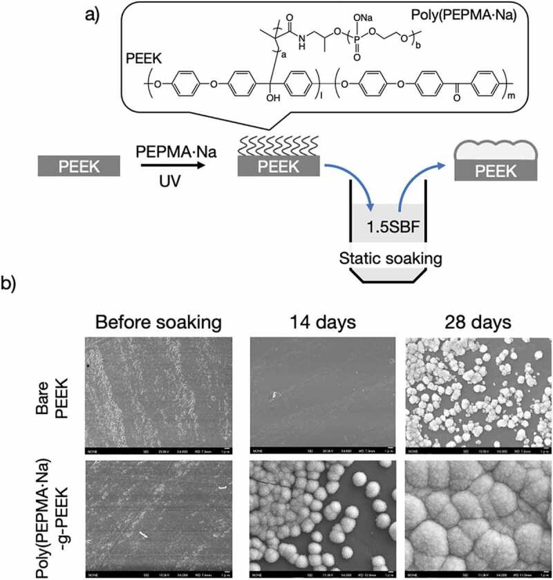
Figure 7.

(a) Schematic of the surface modification of PEEK specimens and (b) SEM images of bare PEEK and PEEK-g-poly(PEPMA·Na) after soaking in x1.5 SBF for 28 days. Reprinted with permission from [93]. Copyright (2019) Taylor & Francis. PEEK, poly(ether ether ketone); SEM, scanning electron microscopy; PEPMA·Na, phosphodeister macromonomers; SBF, simulated body fluid
