Fig. 2.
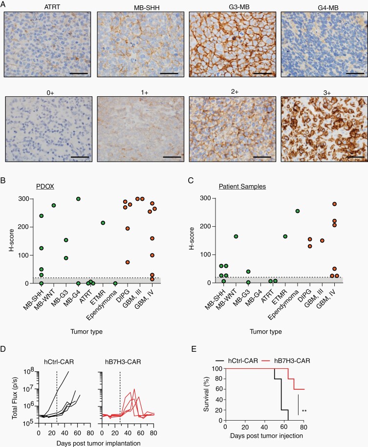
B7-H3 is expressed in pediatric brain tumors. PDOXs were evaluated for B7-H3 expression by IHC. A, Top panel: representative images for different tumor subtypes, including ATRT, MB-SHH, G3-MB, and G4-MB stained for B7-H3. Bottom panel: B7-H3 staining-intensity scale: 0+: no staining, 1+: weak positive, 2+: moderate positive, 3+: strong positive. Scare bar, 50 µm. B and C, H-scores of PDOXs and pediatric brain tumor sections (evaluated by pathologist blinded to tumor type and expression status). D, MB-SHH PDOX tumor cells (Model No. 5, SJMBSHH-13-5634) expressing YFP.ffLuc were implanted into the cortices of NSG mice followed by i.v. injection of 5 × 10⁶ hB7-H3− or hCtrl-CAR T cells on Day 28. Radiance (total flux) is shown. E, Kaplan-Meier survival analysis. Log-rank (Mantel-Cox) test; hCtrl− vs hB7-H3-CAR T cells; P = .0052).

