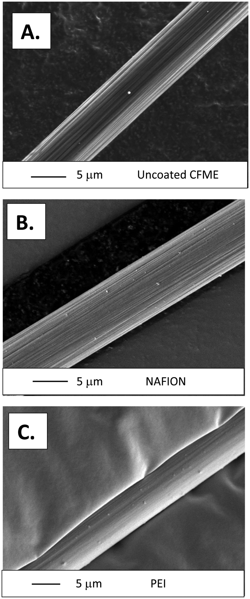
Figure 2:

SEM Images of A). A bare uncoated carbon fiber approximately 7 microns in diameter. B). A carbon fiber electrodeposited in a Nafion solution for approximately 5 minutes. A thin layer of polymer evenly coats the surface of the electrode. C). Polyethyleneimine (PEI) Coated Carbon fibers. The disappearance of the ridges in Figures 2B and 2C indicate that the carbon fibers have been coated in a thin layer of polymer. Small dots on the surface of the fibers are remnants of gold sputtering that were utilized to increase the conductivity, and hence resolution, of the images.
