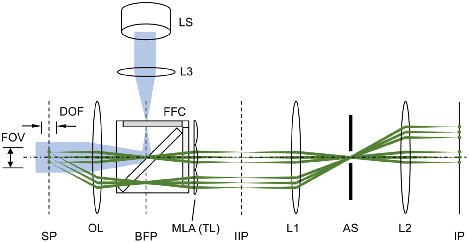
FIG. 1.

Snapshot projection optical tomography (SPOT). The excitation beam path from the light source is shown in blue, and the example emission paths from three fluorophores in the sample plane are shown in green. SP: sample plane; BFP: back focal plane of the objective lens; IIP: intermediate image plane; IP: image plane; OL: objective lens; MLA: microlens array; TL: tube lens; AS: aperture stop; LS: light source; L1, L2, and L3: lenses; FFC: fluorescence filter cube; FOV: field of view; and DOF: depth of field. The dash-dot line represents the optical axis of objective lens.
