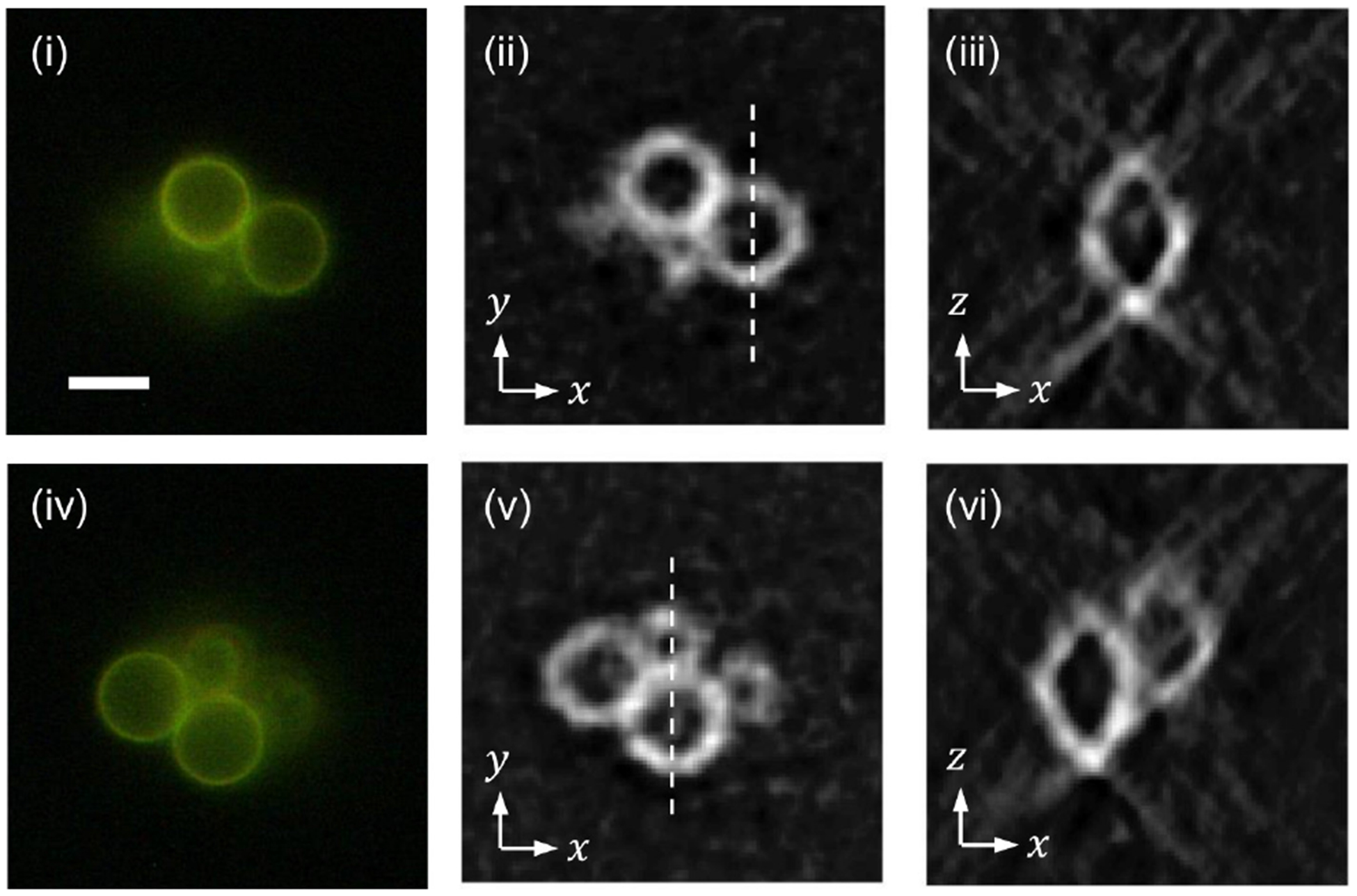
FIG. 8.

Snapshot 3D imaging of a cluster of 6-μm polystyrene beads with the outermost layer stained with fluorescent dye.(i) and (iv): wide-fluorescence images at two axial locations 3 μm apart. (ii) and (v): horizontal cross-sections at the corresponding heights. (iii) and (vi): vertical cross-sections along the dotted lines in (ii) and (v), respectively. The scale bar in (i), 5 μm, applies to all the other images.
