Figure 3.
Stage- and cell-type specific structural plasticity in basolateral amygdala (BLA) inputs to nucleus accumbens (NAc)
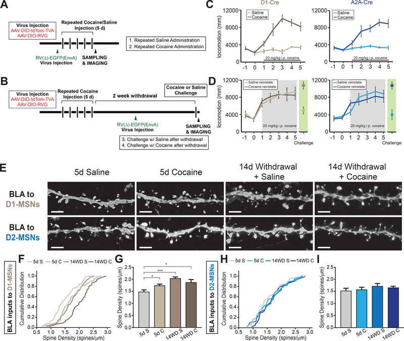
(A and B) Experimental timeline of viral injections and cocaine/saline administrations for 5 day administration group (A) and 5 day cocaine with 2 week withdrawal followed by either saline or cocaine reinstatement injection (B).
(C and D) Both genotypes exhibit locomotor sensitization with successive injections of cocaine (C) that persists after 2 weeks withdrawal with a cocaine, but not saline, reinstatement shot (D).
(E) Representative confocal images of dendritic spines of BLA neurons projecting to D1- and D2-receptor expressing medium spiny neurons (D1-MSNs, D2-MSNs, respectively) in NAc at separate stages of cocaine administration. Scale: 1 μm.
(F) Cumulative distribution plot of spine density of all dendrites imaged in BLA projecting to D1-MSNs. n = 48, 50, 45, 48 dendrites for 5d S (5 mice), 5d C (5 mice), 14WD S (6 mice), 14WD C (6 mice) groups, respectively. p < .05 for 5d S – 5d C and 5d S – 14WD C. p < .001 for 5d S – 14WD S; K-S test.
(G) Average spine densities at separate stages of cocaine administration in BLA dendrites projecting to D1-MSNs. K-S test, dendrites sampled as in (F).
(H) Cumulative distribution plot of spine density of all dendrites imaged in BLA projecting to D2-MSNs. n = 47, 47, 35, 44 dendrites for 5d S (4 mice), 5d C (5 mice), 14WD S (5 mice), 14WD C (6 mice), respectively.
(I) Average spine densities at separate stages of cocaine administration in D2-MSN projecting BLA neuronal dendrites. K-S test, dendrites sampled as in (H).
All data in (G and I) presented as mean ± SEM. Abbreviations: 5d S, 5 day acute saline; 5d C, 5 day acute cocaine; 14WD S, 14 day withdrawal saline reinstatement; 14WD C, 14 day withdrawal cocaine reinstatement. K-S test * P < .05, *** P < .001.

