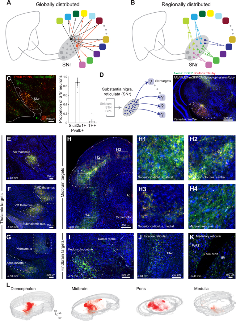
Figure 1. Brain-wide axonal projection pattern from SNr.

(A) Schematic of potential global architecture where intermixed SNr neurons project to all target regions with equal probability, although not necessarily equal axonal density.
(B) Schematic of potential regionally divided architecture where different pools of neurons project to each target and, further, the projection pools are spatially segregated.
(C) Representative in situ hybridization of Pvalb (red) and Slc32a1 (green) mRNA in SNr (left). Proportion of Pvalb+ GABAergic and Th+ dopaminergic neurons in SNr (right, 39 sections). Error bars = SEM.
(D) Anterograde tracing of SNr projections labeled with AAV-DJ-hSyn-FLEX-mGFP-2A-Synaptophysin-mRuby injected into SNr of a PV-Cre mouse.
(E-K) Whole-brain, high-resolution (0.325 μm/pixel) slide scanning of SNr axons (green) and boutons (red) demonstrates projections to (panels E-G) motor and intralaminar/midline thalamus, subthalamic, and incertal nuclei, (panels H1-H3) domains of the superior colliculus, and (panels H4, I-K) diverse premotor brainstem regions, including midbrain, pontine, and medullary reticular formations.
(L) Spatial distribution of Synaptophysin-mRuby labeled SNr boutons in 3D reconstructions of the diencephalon and general divisions of the brainstem. See also Figure S1.
