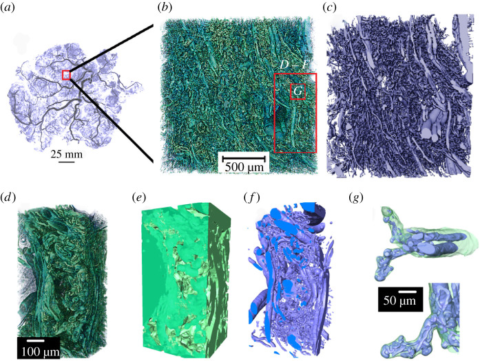
Figure 1.
Multi-scale tissue architecture of placental tissue from synchrotron micro-CT. (a) Placental cast under micro-CT (unpublished image from [5]). (b–g) Images demonstrating the complex hierarchical architecture of the human placenta (Specimen 1, normal placenta at term). (b) Three-dimensional rendering of approximately 8 mm3 human placental tissue. (c) Fetal vascular network segmented from placental tissue using a U-Net algorithm. (d–f) A small section of the placental tissue was cropped from the original dataset (red box in b) and 3D rendered. (d) Three-dimensional rendering of approximately 0.2 mm3 tissue showing different hierarchical features. (e) U-Net segmented fetal tissue component, (f) fetal vessels and (g) fetal capillary network with surrounding villous tissue overlaid.

