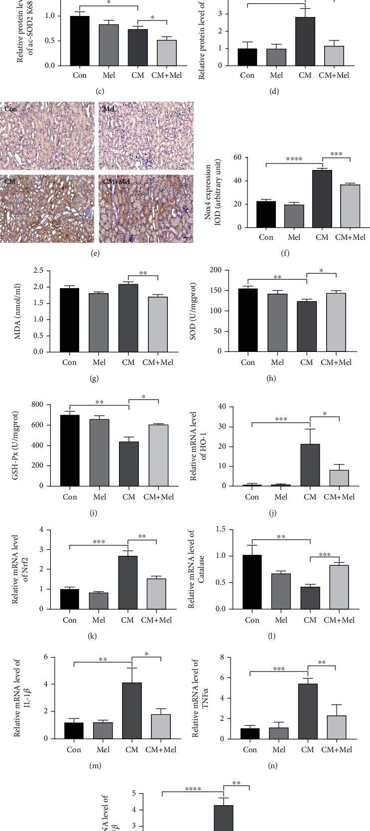
Figure 2.

Melatonin decreased kidney oxidative stress and inflammation caused by iohexol in vivo. (a) Representative western blots. (b–d) Quantitative analysis of protein expression of Sirt3, ac-SOD2 K68, and Nox4. (e) Representative immunohistochemical staining of Nox4. (f) Quantitative analysis of Nox4 immunostaining. (g) Renal MDA content. (h) Renal SOD activity. (i) Renal GSH-Px activity. (j–l) Quantitative analysis of HO-1, Nrf2, and Catalase mRNA expression in the kidney of four groups. (m–o) Quantitative analysis of IL-1β, TNFα, and TGFβ mRNA expression in the kidney of four groups. MDA: malondialdehyde. SOD: superoxide dismutase. GSH-Px: glutathione peroxidase. All experiments were repeated at least 3 times. Data are expressed as mean ± SEM. ∗P < 0.05, ∗∗P < 0.01, ∗∗∗P < 0.001, ∗∗∗∗P < 0.001.
