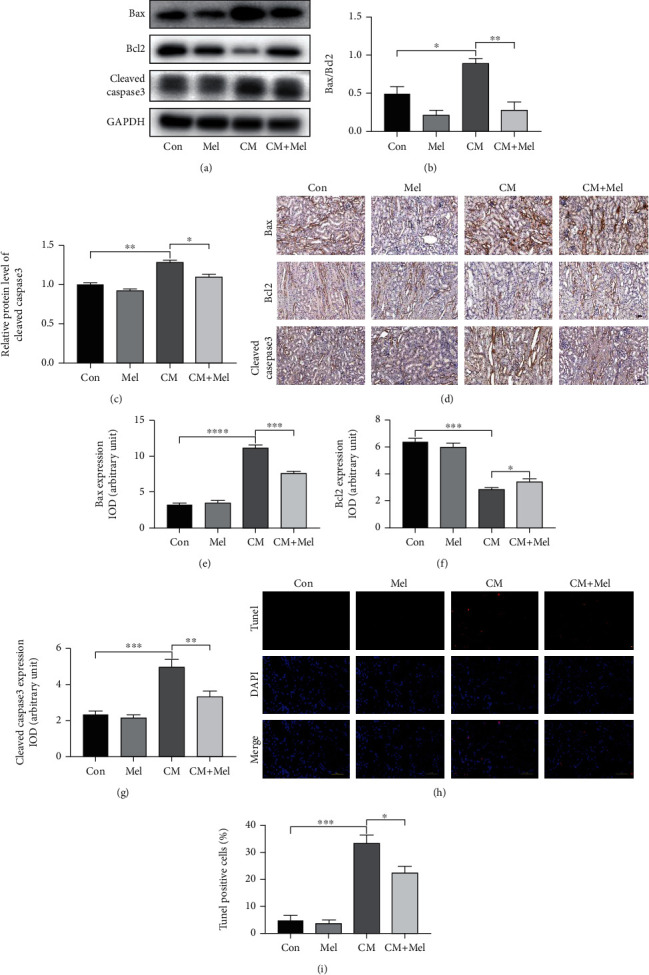
Figure 3.

Effect of melatonin treatment on antiapoptosis after iohexol administration in vivo. (a) Representative western blots. (b, c) Quantitative analysis of the ratio of Bax to Bcl-2 and the expression of cleaved caspase3 were examined. (d) Representative immunohistochemical staining of Bax, Bcl2, and cleaved caspase3. (e–g) Quantification of Bax, Bcl2, and cleaved caspase3. (h) Representative digital images of apoptotic cells. The apoptotic cells were detected by TUNEL (red), and the nuclei were detected by DAPI (blue). (i) Percentage of TUNEL-positive nuclei. All experiments were repeated at least 3 times. Data are presented as mean ± SEM. ∗P < 0.05, ∗∗P < 0.01, ∗∗∗P < 0.001, ∗∗∗∗P < 0.001.
