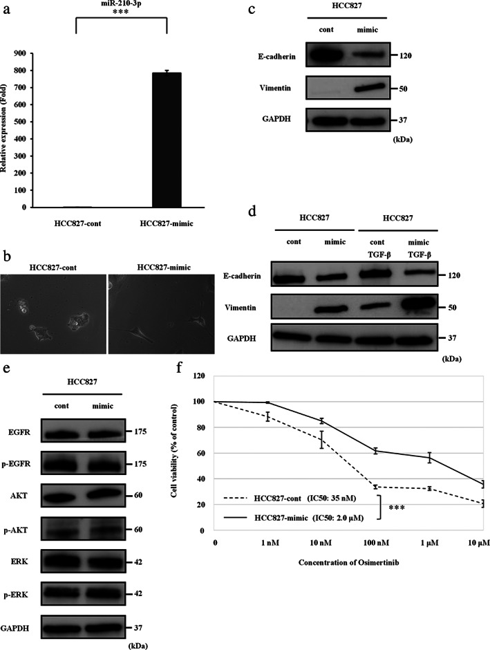
FIGURE 5.

MiR‐210‐3p promotes EMT and resistance to osimertinib in HCC827 cells. (a) MiR‐210‐3p expression in HCC827 cells after transfection of the miR‐210‐3p mimic, as determined by qRT‐PCR analysis. Control mimic (cont); miR‐210‐3p mimic (mimic). Data are expressed as the mean ± SD from three independent experiments. ***p < 0.001. (b) HCC827‐cont cells and HCC827‐mimic cells observed under a light microscope. (c) Protein levels of EMT markers in HCC827 cells after transfection of miR‐210‐3p mimic, as shown by western blotting analysis. (d) Protein levels of EMT markers in HCC827 cells after transfection of the miR‐210‐3p mimic with or without TGF‐β stimulation. (e) Protein expression of EGFR signaling pathway molecules in HCC827 cells after transfection of the miR‐210‐3p mimic. (f) The results of the cell viability assays are shown. Data are expressed as the mean ± SE from three independent experiments. ***p < 0.001. EGFR, epidermal growth factor receptor; EMT, epithelial–mesenchymal transition; ERK, extracellular signal‐regulated kinase; GAPDH, glyceraldehyde‐3‐phosphate dehydrogenase; IC50, concentration needed for 50% reduction of the growth; p‐EGFR, phosphorylated‐EGFR; qRT‐PCR, quantitative reverse transcription‐polymerase chain reaction; SD, standard deviation; SE, standard error; TGF‐β, transforming growth factor‐β
