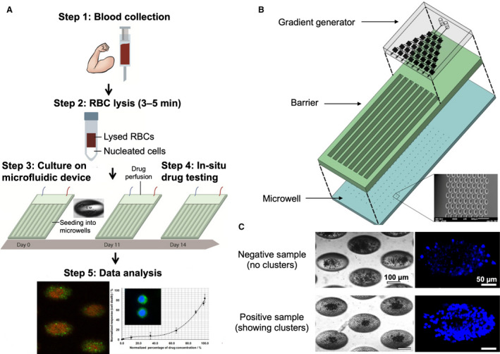
Fig. 3.

CTC cluster assay for drug screening. (A) Schematic depicting the cluster assay for anticancer drug screening. Blood samples are first lysed to remove red blood cells (RBCs), and the nucleated cell fraction is then seeded into an integrated microwell‐based microfluidic device. Drugs are introduced directly in situ, and the CTC clusters are generated within 2 weeks. (B) Three‐dimensional layout of the drug assay device showing the gradient generator, barrier, and microwells. (C) Representative bright‐field and fluorescent images of negative and positive samples. Nuclei were stained using Hoechst dye. Figures adapted with permission from Ref. [105].
