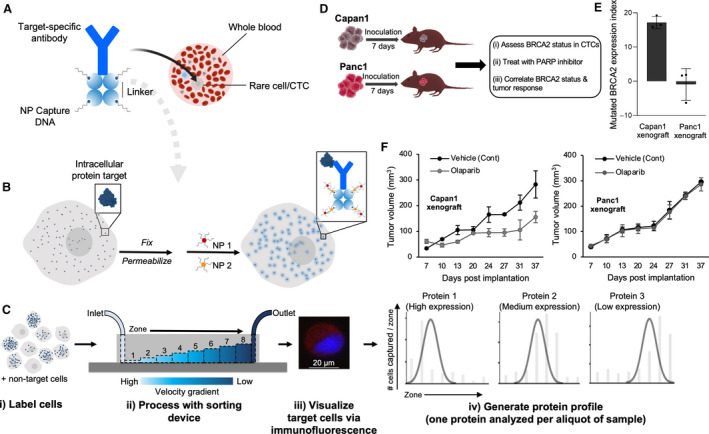
Fig. 4.

Analysis of intracellular proteins using the MagRC approach. (A) An antibody specific for the target intracellular protein is tagged with streptavidin then modified with biotin‐labeled ssDNAs using biotin–streptavidin coupling. (B) The cells expressing the target intracellular protein are fixed, permeabilized, and incubated with the intracellular protein‐specific antibody modified with ssDNAs. The ssDNAs are then hybridized with two capture probes (CP1 and CP2), which are composed of complementary DNA sequences modified at one end with MNPs. Aggregates of MNPs are thus formed and trapped within the cells that express the intracellular protein. (C) The cells are sorted using a microfluidic device featuring eight capture zones, immunostained, and counted to generate a profile characteristic for the target protein. (D) Schematic illustration for therapeutic protein analysis in xenografted mice. (E) Analysis of mutated BRCA2 protein in CTCs captured from the blood of mice bearing either Capan1 (with mutated BRCA2) or Panc1 (with wild‐type BRCA2) xenograft. The analysis was carried out at day 7 after tumor formation. After tumor formation, the mice were randomly divided into control and treated groups. Mice in the treated group received 50 mg·kg−1 olaparib, whereas mice in the control group received only the vehicle. (F) Tumor volume is plotted against duration of treatment for Capan1 and Panc1 xenografts. Figure reprinted with permission from Ref. [54].
