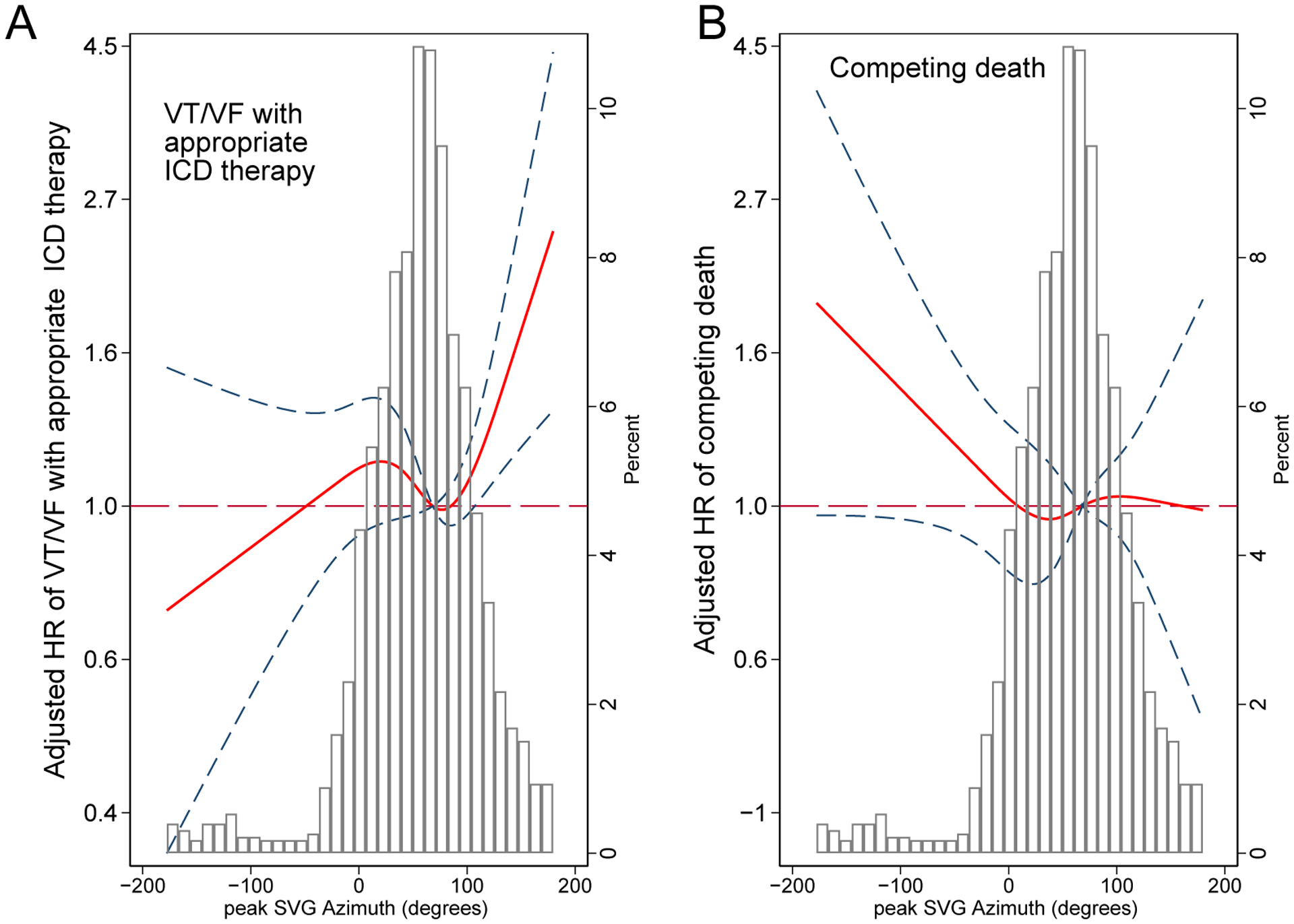
Figure 4.

Adjusted (model 1) risk of (A) sustained VT/VF with appropriate ICD therapy and (B) competing death without appropriate ICD therapy associated with peak SVG azimuth. Restricted cubic spline with 95% CI shows a change in cause-specific Cox HR (Y-axis) in response to peak SVG azimuth change (X-axis). The 50th percentile of the SVG azimuth is a reference. Knots of the peak SVG azimuth are at (-21)-42-75-137 degrees.
