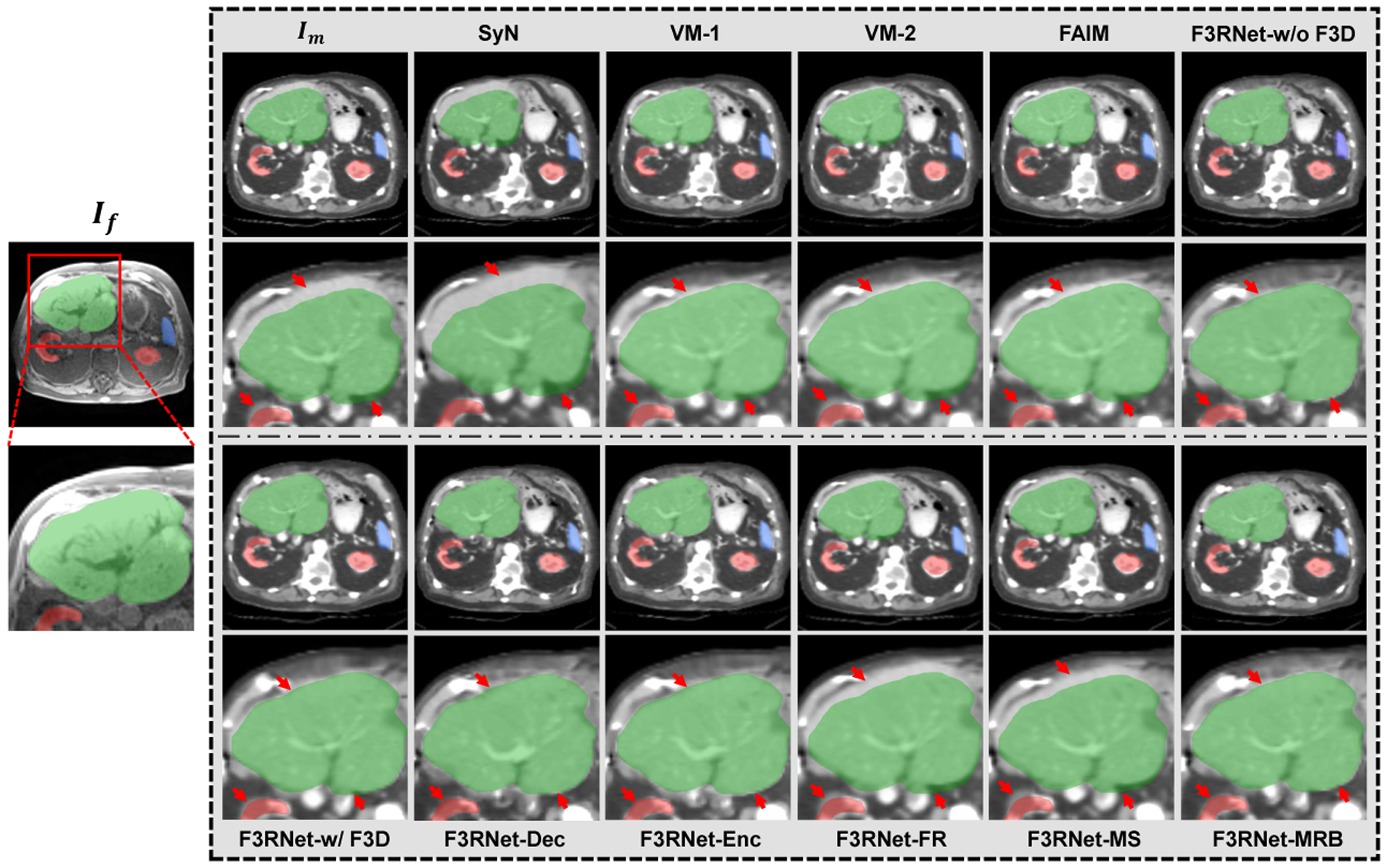
Fig. 4.

Visual results of an example for CT-to-MRI registration. Outside the grey box shows an example fixed MR image and a zoom-in region with the segmentation masks of the liver (green), kidney (red), and spleen (blue). The corresponding warped CT images and zoom-in regions for baselines and ablation study are presented in the grey box. A good registration will cause structures in warped images to close to the corresponding fixed segmentation masks. The red arrows indicate the registration of interest at the boundary of the organ.
