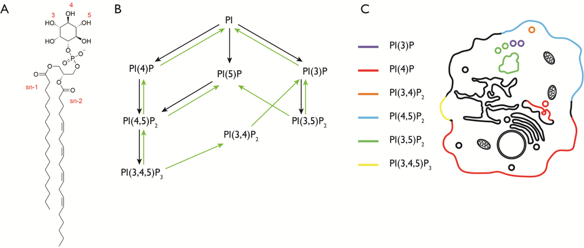
Figure 1. Structures and subcellular localization of the phosphoinositides.

(A) The structure of phosphatidylinositol (PI). The hydroxyl groups of the inositol ring are phosphorylated at the indicated positions (3, 4, and/or 5) to generate phosphoinositides (PIPs). The illustrated PI structure is the most abundant in mammalian cells, containing stearoyl and arachidonyl groups at the sn-1 and sn-2 positions respectively. (B) The seven different PIPs are shown, with known pathways of conversion from one species to another. Black arrows represent phosphorylation reactions and green arrows represent dephosphorylation reactions. (C) Cartoon illustrating the localization of the major pools of the seven different PIP species.
