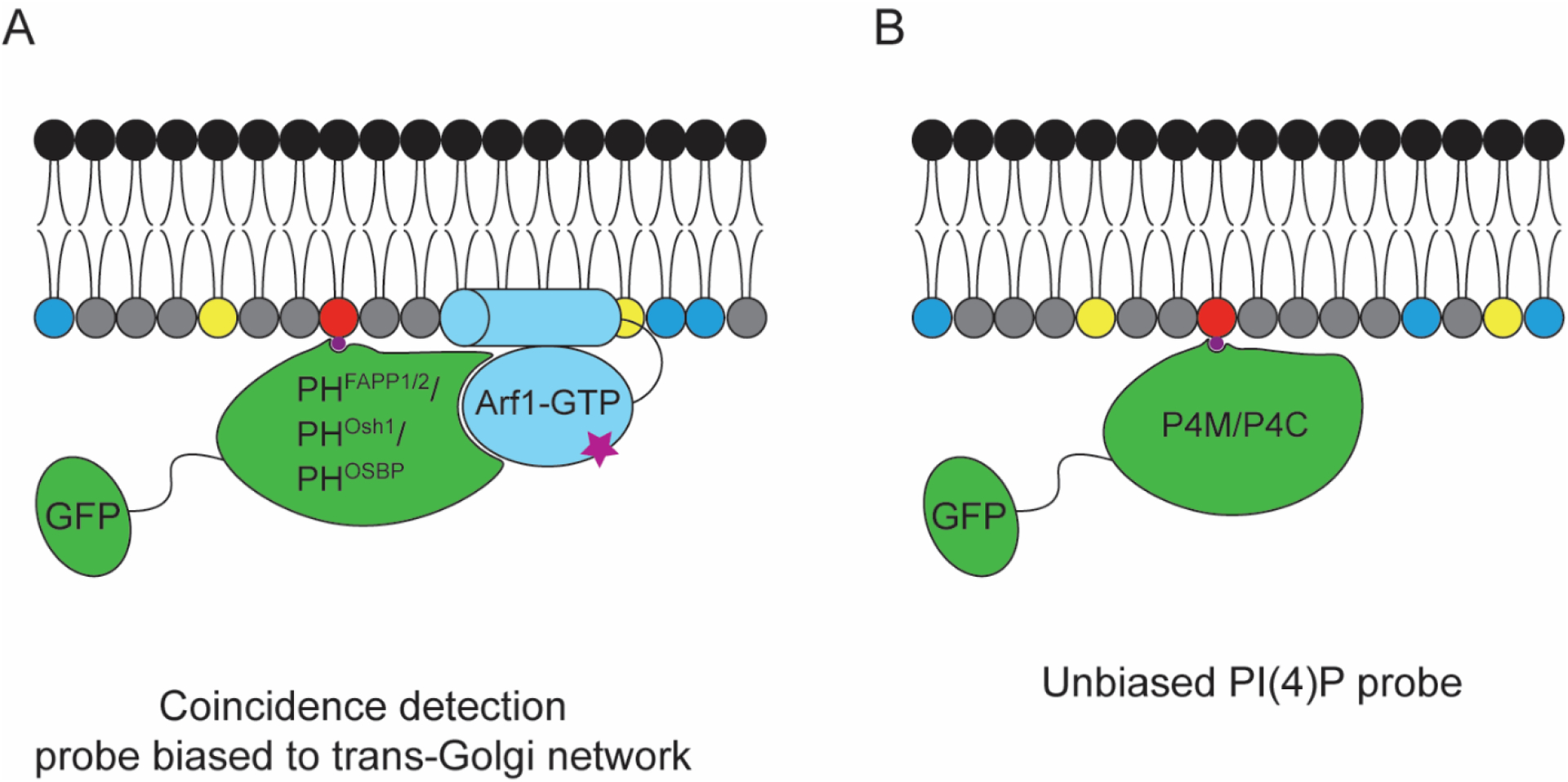
Figure 4. Genetically encoded probes for visualizing PI(4)P involving fluorescent protein fusions to PI(4)P-binding domains.

(A) PH domain-based probes from FAPP1, Osh1, and OSBP, capable of recognizing PI(4)P, but which are typically biased toward Golgi localization due to a coincidence detection mechanisms via secondary interactions with Arf1. (B) Unbiased PI(4)P probes take advantage of PI(4)P-binding motifs from Legionella pneumophila proteins SidM and SidC, termed P4M and P4C respectively, that do not engage in additional interactions that might affect probe localization.
