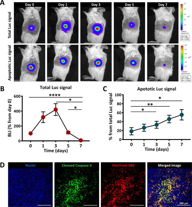
Fig. 4. In vivo apoptosis of MSC after intrapancreatic transplantation.
A Representative BLI time-course images obtained after injection of D-Luciferin (for detection of total viable cells) or Z-DEVD-aminoluciferin (for detection of apoptotic cells), after intrapancreatic transplantation of Luc+ MSC; B Quantification of BLI signal measured after D-Luciferin administration in mice; data are shown as mean ± SEM (n = 5); C Quantification of BLI signal measured after Z-DEVD-aminoluciferin administration in mice; data are shown as mean ± SEM (n = 5). D Immunofluorescence images of the pancreas of a NOD mouse at 7 days after MSC local transplantation, indicating cellular apoptosis at the site of transplantation. Note that numerous VT680-labbeled MSC were apoptotic at that time, as revealed by the presence of cells co-stained with VT680 (red pseudo-colour) and cleaved Caspase 3 (green pseudo-color).

