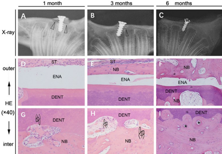
Fig. 6.
A–C A graft heals with the original bone, and the low-density region represented by the triangles which gradually disappears. Histologically, D the enamel layer is covered with soft tissue, E with a thin layer of new bone, and F with newly remodeled bone on the surface of enamel. G–I Around dentin, vascularization and osteoclast-like cells (finger mark) and new bone formation are observed with bony lacunae (asterisk) which indicates bone remodeling process [72]. Reproduced with permission of DENTAL TRAUMATOLOGY *DENT dentin of tooth, ENA enamel of tooth, NB new bone, ST soft tissue

