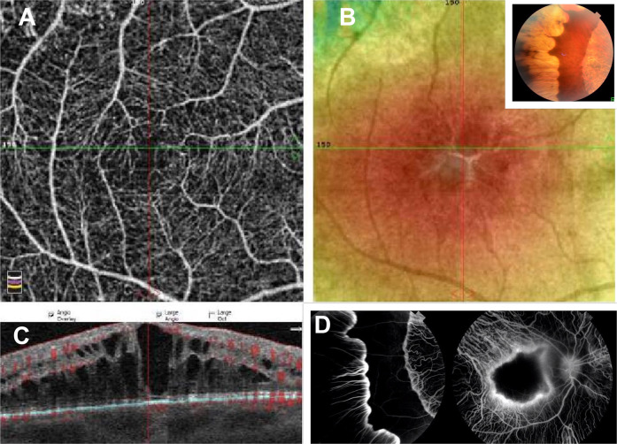
Fig. 1. A composite of the right posterior pole in a 45-year-old Caucasian woman with best spectacle-corrected visual acuity of 6/21 (20/70) and −4D myopia.
The superficial foveal avascular zone (a) is very small on 3 × 3 mm OCTA map. The superficial and capillary plexuses were well preserved with no focal losses. Fundus photograph of the macula (b) shows prominent cystoid spaces well delineated on linear OCT scan (c). Autofluorescence shows sparing of the macula. The inset (b) shows the classical scalloped areas of chorioretinal degeneration advancing from the periphery. Fluorescein transits of the equator and macula (d) shows diffuse chorioretinal atrophy sparing a sector of the midperiphery and macula.

