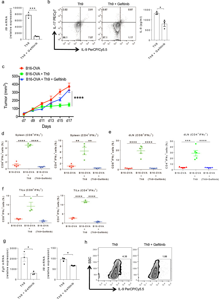
Fig. 2. EGFR inhibition abrogates the anti-tumor functions of Th9 cells.
a, b Naïve CD4+ T cells from WT mice were in vitro differentiated under Th9 conditions with or without 1.0 μM gefitinib for 3 days followed by a. qPCR analysis of Il9 expression. b ELISA for IL-9 and flow cytometry analysis of intracellular staining for IL-9 and IL-17. Data are representative of mean ± SEM from three independent experiments. c–f Naïve CD4+ T cells from OT-II TCR transgenic mice were in vitro differentiated into Th9 with or without 1.0 μM gefitinib for 3 days. Cells were then adoptively transferred into B16-OVA tumor-bearing WT mice, randomized into three groups (n = 5 mice per group). c Mean tumor volume was measured over time shown as tumor growth curve. d, e Spleen and tumor draining lymph nodes (dLN) were harvested and single cell suspensions were made followed by FACS analysis of intracellular staining for CD4+IFNγ+ and CD8+IFNγ+. f TILs were isolated from the tumor followed by FACS analysis of intracellular staining for CD8+IFNγ+and CD4+IFNγ+ cell populations. Data are representative of mean ± SEM from three independent experiments. g, h Sorted naïve human CD4+ T cells were differentiated into Th9 cells with or without 1.0 μM gefitinib. g mRNA expression of Egfr and Il9 was determined by qPCR. Data are representative of mean ± SEM from three healthy individuals. h Intracellular staining for IL-9. a, b ***P = 0.0003, *P = 0.017, using two-tailed unpaired Student’s t test. c ****P < 0.0001, using two-way ANOVA followed by Tukey’s multiple comparison test. d–f ****P < 0.0001, **P = 0.001, ***P = 0.0001, *P = 0.02, using one-way ANOVA followed by Tukey’s multiple comparison test. g *P = 0.014, using two-tailed unpaired Student’s t test.

