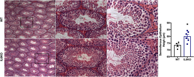
Figure 1.
Testicular morphometric and histological analyses: WT group (a) and IL6KO group (b). Thicker layer of the seminiferous epithelium in the testes of IL6KO mice (p = 0.0381) without apparent morphological alteration. *p < 0.05, Mann–Whitney test. Values expressed as mean ± SEM. Results are representative of samples from 4 to 6 mice per group. The images show the seminiferous tubule under obj.5x, obj.20 × and obj.40× magnifications (from left to right, respectively). Ep, seminiferous epithelium; Lu, lumen; Is, interstitial space. Black rectangle (left panel) shows the seminiferous tubule in the middle panel under high magnification. White dotted line (middle panel) indicates the seminiferous epithelium thickness.

