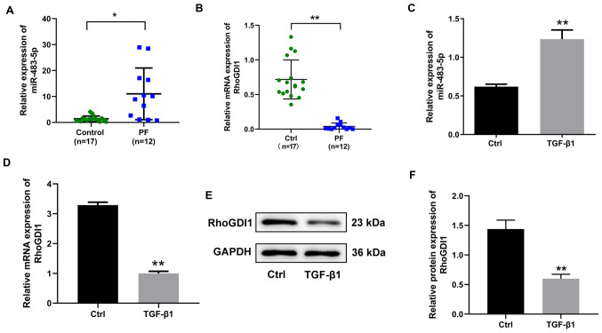
Figure 1.
Expression of miR-483-5p and RhoGDI1 in human PF tissue and A549 cells treated with TGF-β1. The relative expression of (A) miR-483-5p and (B) RhoGDI1in PF (n=12) and normal lung tissue (n=17) was determined by RT-qPCR. *P<0.05 and **P<0.01. (C) Relative expression of miR-483-5p in A549 cells treated with 10 ng/ml TGF-β1 for 48 h was determined by RT-qPCR. The relative (D) mRNA and (E and F) protein expression of RhoGDI1 in A549 cells treated with 10 ng/ml TGF-β1 for 48 h were detected by RT-qPCR and western blot. Data are presented as the mean ± SD (n=3). **P<0.01 vs. Ctrl. PF, pulmonary fibrosis; RhoGDI1, Rho GDP dissociation inhibitor 1; RT-qPCR, reverse transcription-quantitative PCR; TGF-β1, transforming growth factor-β1; miR, microRNA; Ctrl, control.

