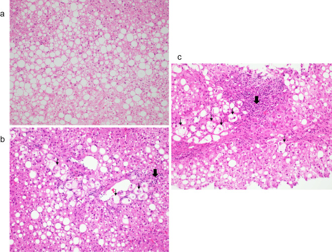
Figure 2.
Hematoxylin and Eosin staining of liver biopsy tissues at the initial presentation (a), after 12 years (b), and after 14 years (c) (×40). (a) Approximately 80% of the tissues show fat deposits and macrovesicles to medium-sized fat droplets. No hepatocyte balloon-like cells are observed (grade 0). (b) Fatty deposits in the liver are decreased to approximately 40% of the tissue. Lipid droplets, ballooning, and Mallory bodies are also observed. Inflammatory cell infiltrates, mainly comprising lymphocytes, are distributed in the portal region (grade 2). (c) Fatty deposits in the liver comprise approximately 40% of the tissue. Lipid droplets, ballooning, and Mallory bodies are also observed. Inflammatory cell infiltrates are distributed in the portal area (grade 2). (↓) Inflammatory cell, (⬇) Ballooning cell

