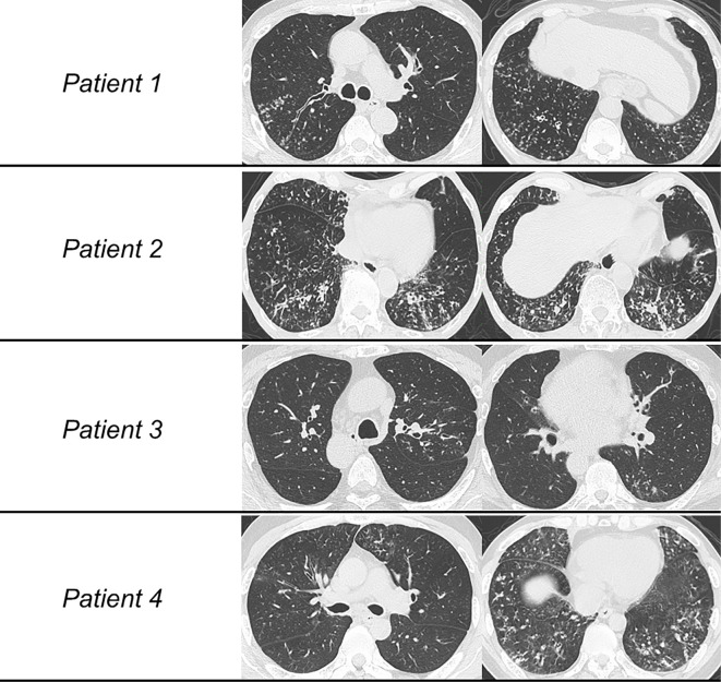
Figure 1.
High-resolution computed tomography (HRCT) images of the four patients. (Patient 1) Centrilobular nodular shadows and bronchial thickening are diffusely distributed predominantly in the lower lungs. (Patient 2) HRCT showed not only diffuse centrilobular nodules but also mosaic perfusion indicative of air trapping and mild consolidation. (Patient 3) Centrilobular nodules and bronchiectasis were seen predominantly in the dorsal left upper lung and left S6. (Patient 4) Diffuse distribution of centrilobular nodules, bronchial thickening, and air trapping were seen predominantly in the lower lungs.

