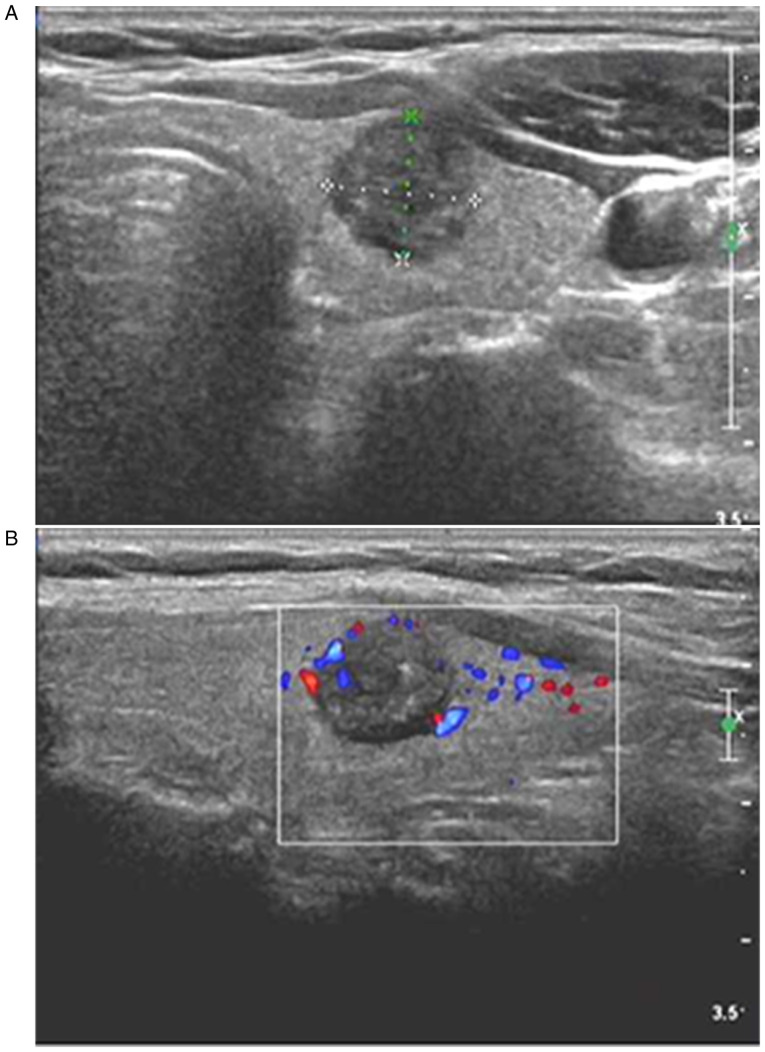
Figure 1.
Diagnosis of MTC in a 42-year-old women through postoperative pathology. (A) Two-dimensional ultrasound showing a solitary nodule in the upper pole of the right lobe of thyroid gland, with an oval shape, an aspect ratio of <1, a clear boundary, hypoecho, and internal coarse calcification. (B) CDFI showing blood flow signals inside and around the nodule; the ultrasound results suggested that the nodule met the TI-RADS category 5, and that it may be MTC. MTC, medullary thyroid carcinoma; CDFI, color Doppler flow imaging.

