Figure 3.
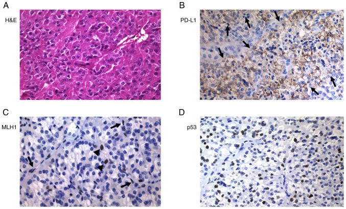
Poorly differentiated thyroid carcinoma (Hürthle cell variant; case 19). (A) H&E staining. (B) Strong positivity for PD-L1 (arrows). (C) Tumor cells were negative for MLH1, but positivity was observed in stromal cells, endothelial cells (arrows) and lymphocytes (arrowheads) (internal control). (D) Strong positivity for p53. Magnification, ×400. H&E, hematoxylin and eosin; PD-L1, programmed death ligand 1; MLH1, mutL homolog 1.

