Figure 5.
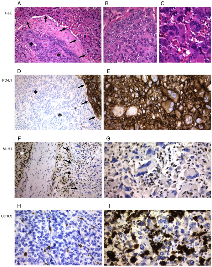
ATC with poorly differentiated areas (case 15). (A) Transition between poorly differentiated areas (asterisks) and ATC areas (arrows) (magnification, ×200). Morphological differences between (B) poorly differentiated and (C) ATC areas at a higher magnification (magnification, ×400). (D) PD-L1 expression was only detected in the ATC areas (arrows), but not in the poorly differentiated component (asterisks) (magnification, ×200). (E) ATC cells showed strong and diffuse positivity for PD-L1 (magnification, ×400). MLH1 expression at magnification (F) ×200 and (G) ×400 was found in the poorly differentiated areas (asterisk), but not in the ATC areas (arrows). CD163+ macrophages were not detected in (H) the poorly differentiated areas, but were heavily detected in (I) the ATC areas (magnification, ×400). ATC, anaplastic thyroid carcinoma; H&E, hematoxylin and eosin; PD-L1, programmed death ligand 1; MLH1, mutL homolog 1.

