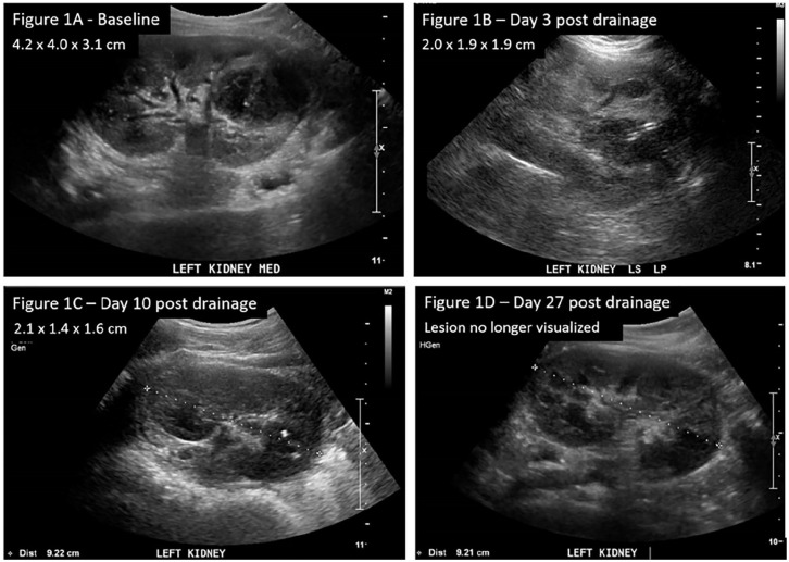
Figure 1.
Ultrasound of kidneys, ureters, and bladder. (A) (Baseline) A heterogeneous structure noted in the left kidney extending from the mid to lower pole. This lesion is consistent with a renal abscess and much of it appears liquefied. (B) (Day 3 post drainage) Abscess decreased in size. (C) (Day 10 post drainage) Previously noted abscess smaller in size with catheter in situ. (D) (Day 27 post-drainage) Cystic lesion no longer visualized.

