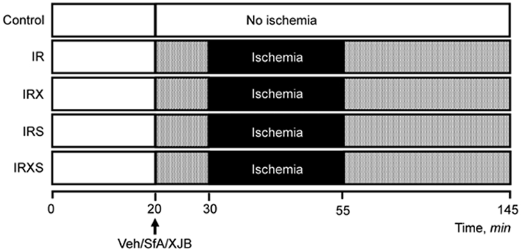
Fig. 1.

Design of experiments. Groups: i) control, non-ischemic hearts perfused continuously for 145-min (n=7); ii) IR, hearts subjected to 25-min ischemia followed by 90-min reperfusion (n=6); iii) IRX, hearts subjected to 25-min ischemia followed by 90-min reperfusion in the presence of 0.2 μM XJB (n=6); iv) IRS, hearts subjected to 25-min ischemia followed by 90-min reperfusion in the presence of 0.5 μM SfA (n=7); and v) IRXS, hearts subjected to 25-min ischemia followed by 90-min reperfusion in the presence of XJB and SfA (n=7). Details are given in Materials and Methods.
