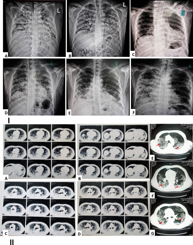
Fig. 1.
(I) Chest X-ray image of a COVID-19 positive old male, showing bilateral mid and lower zones homogenous consolidation in peripheral distribution along with Obscuration of both CP angles (red arrow), findings fall in the category of indeterminate for COVID-19 (A–F). (II) Axial thin-section chest computed tomography (CT) in patients with COVID-19 pneumonia from our institution. A–G Non-contrast chest CT scan exam showed bilateral ground glass opacities and interlobular septal thickening giving appearance of crazy paving and areas of surrounding consolidation giving appearance of reverse halo sign and air bronchogram (red arrow) (Color figure online)

