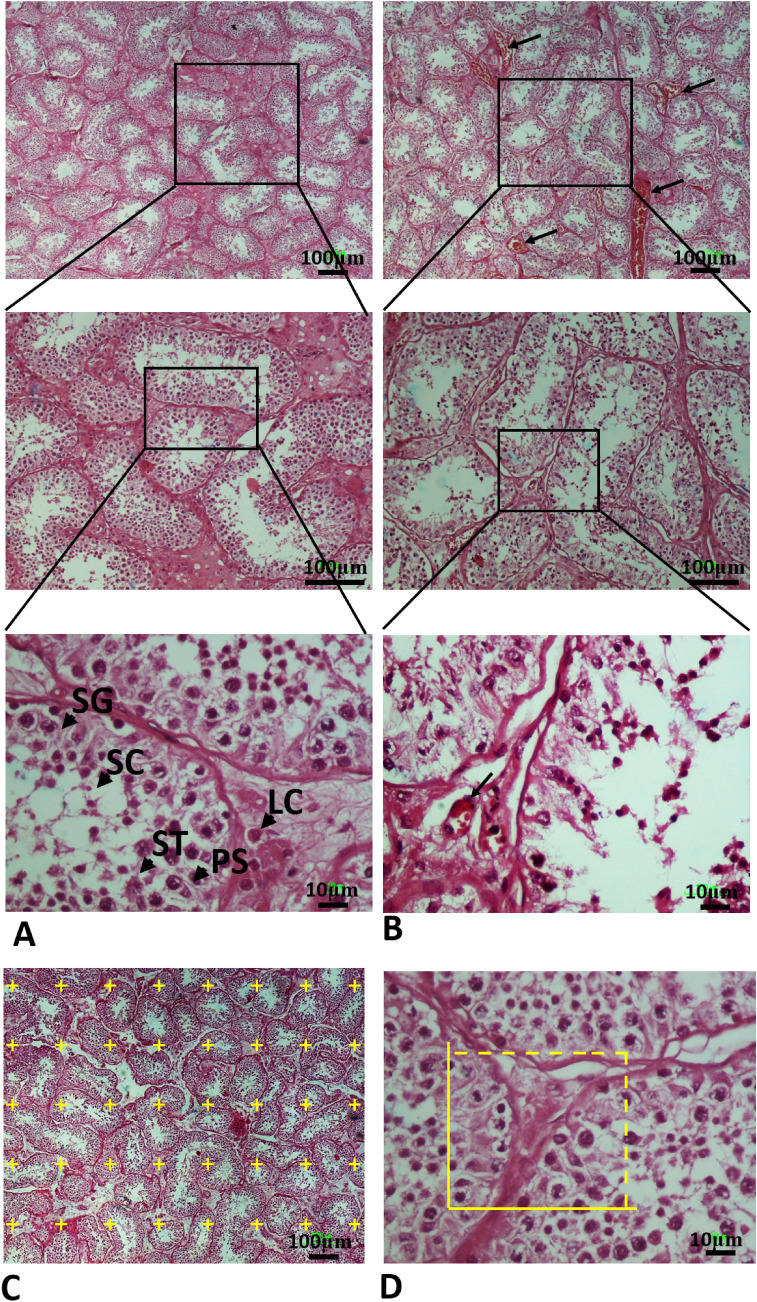
Fig. 3.
Photomicrograph of the testis stained with H&E; (× 4, × 10 and × 40). SG Spermatogonia (SG), primary spermatocyte (PS), round spermatid (ST), Sertoli cell (SC), Leydig cell (LC). A In the control group, the testis tissue, including the seminiferous tubules and germinal epithelium, has a normal appearance. B The testicular cells death, interstitial edema and thinning of the seminiferous epithelium was observed in all autopsy COVID-19 specimens of testes. C A point grid is superimposed over the photomicrograph for measuring the volume using Cavalieri method. D Counting frames are superimposed over the photomicrographs to measure the number of testicular cells using optical disector method. The yellow box is counting frame (Color figure online)

