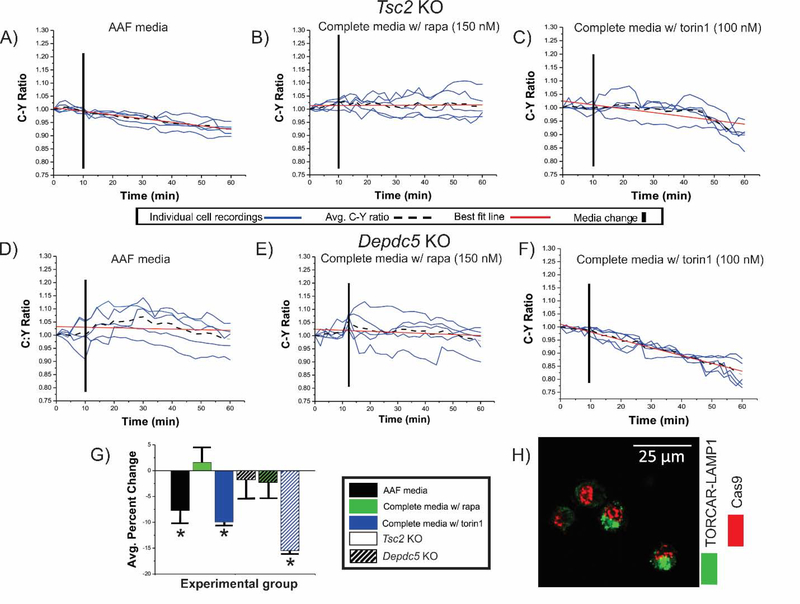
Figure 2: Decrease in C:Y ratio is observed in Tsc2 KO, but not Depdc5 cells after incubation in AAF media.
Tsc2 KO (solid bars in G) and Depdc5 KO (striped bars in G) cells transfected with TORCAR were incubated in AAF media (A, D), in complete media with rapamycin (B,E), or in complete media with torin1 (C,F). After incubation in AAF media (A), C:Y was decreased in Tsc2 KO cells (7.0%, (G); p<0.05). No change in C:Y was observed after incubation in complete media containing rapamycin in Tsc2 KO (B) and Depdc5 KO (E) N2aC, respectively. An average percent decrease in C:Y of 9.9% and 15.4% was observed in both Tsc2 KO and Depdc5 KO N2a cells (p<0.05) after incubation in complete media containing torin1 (C,F), respectively. In (H), representative images of TORCAR and CRISPR/Cas9 plasmid co-transfected cells are shown. AAF= amino acid free, rapa = rapamycin, KO = knockout. Error bars in (D), standard error of the mean. *p<0.05.

