
Fig. 6.
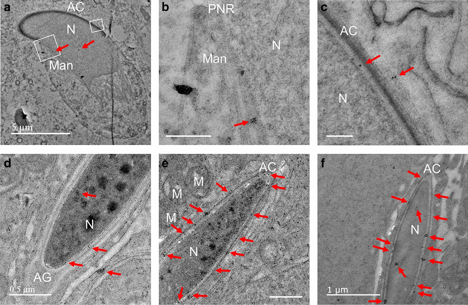
Localization of PRAMEL1 in the acrosome (AC) and manchette (Man). The image in A shows a step 9 spermatid. Clusters of PRAMEL1 gold particles were detected in the manchette (boxed region in A) and the enlarged image in B. A low density PRAMEL1 labeling was seen in the acroplaxome (C) (the boxed region in A). In steps 10–12 spermatids, clusters of gold particles were seen in the nucleus (N) and acrosome region (D, E), but a very few gold particles were observed in the acrosome granule (AG) or acrosome matrix (AM) located in the front head (D, E). In step 16 elongated spermatid, large clusters of gold particles were observed in nucleus and acrosome (F). Some of the clusters were in the nuclear membrane adjacent to acrosome. M: mitochondria; PNR: perinuclear ring; Unlabeled scale bar = 0.5 µm