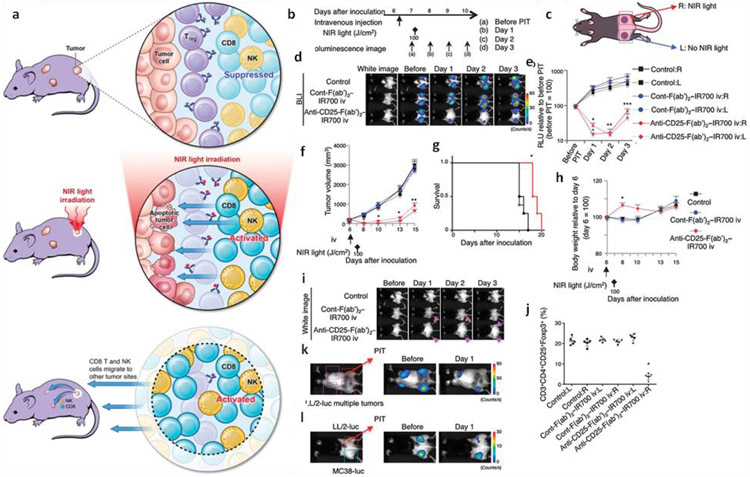
Fig. 6.
a) Schematic representation of the induced immunotherapy after NIR-PIT. b) General regimen adopted for the NIR-PIT. c) Biflanked tumor model: right tumor was treated with NIR light and anti-CD25-F(ab′)2–IR700 or (ab′)2–IR700 driven phototherapy, while the left tumor was left untreated. d) Representative bioluminescence images showing the changes after the localized NIR-PIT. e) Reduction in the relative light units (RLU) of the right tumor after NIR-PIT as well as in the left non-irradiated tumor. f) Reduction in size of the tumor after the CD25 treated right tumor as well as non-irradiated one. g) Prolonged survival of mice after NIR-PIT. h) Change in weight after the NIR-PIT, i) Edema due to NIR-PIT at both tumors after CD25 targeted NIR-PIT, j) Depletion of CD4+CD25+Foxp3+ Tregs in the right tumor but not in the left untreated. k) Regression of multiple LL/2-luc tumors after the localized CD25 targeted NIR-PIT at the right tumor. l) Negligible effect on the MC38-luc tumor after NIR-PIT on the LL/2-luc tumors of the right side. Adopted with permission from reference [124].

