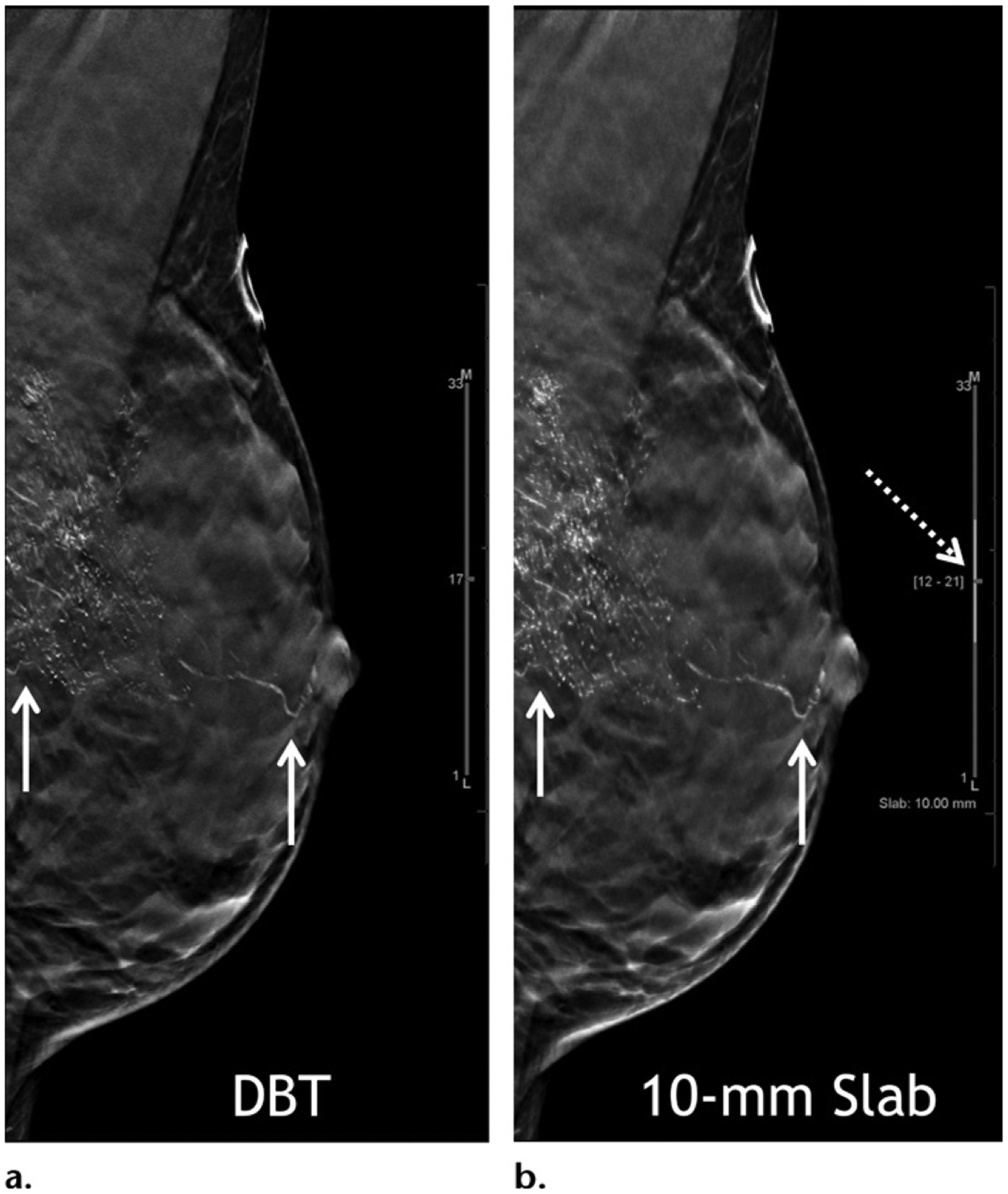
Figure 9.

High-grade ductal carcinoma in situ at baseline screening in a 40-year-old woman. Mediolateral oblique in-plane DBT (a) and 10-mm slab (b) images show segmentally distributed fine linear branching calcifications in the left breast (solid arrows). Currently the standard section thickness for DBT reconstruction images is 1 mm. Slab viewing is possible (dashed arrow in b) with adjustable slab thickness to allow quicker review. In this patient, slab viewing also allows a maximum intensity projection–like capture of the overall extent of the abnormality. Stereotactic biopsy was performed and yielded a diagnosis of high-grade ductal carcinoma in situ.
