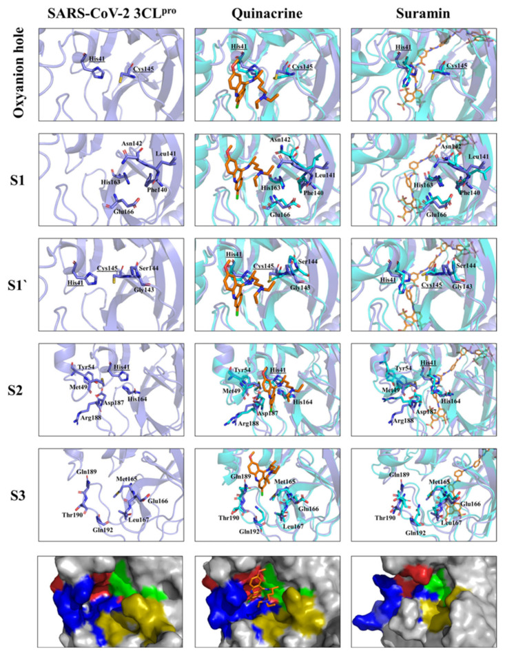
Figure 6.
Substrate-binding pocket and oxyanion hole of SARS-CoV-2 3CLpro (S1, S1’, S2, and S3) free accessibility and under quinacrine and suramin influence. The structural overlay between the single protein and in complex, the active site residues are highlighted. The amino acid residues are shown in sticks. The surface view (zoom) of the substrate-binding area demonstrates the occupied area by the molecules and the conformational changes induced by its binding. Substrate-binding subsites highlighted in green for S1’, gold for S1, red for S2, and blue for S3.

