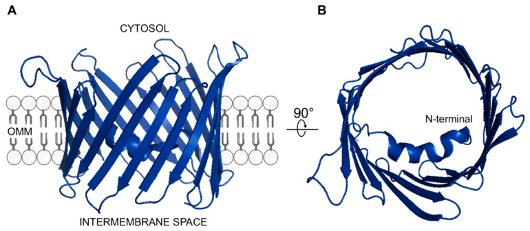
Figure 1.
3D structure of human VDAC1 (A) Lateral view showing the β-barrel organization. The protein is embedded in the OMM facing hydrophilic residues towards cytosolic or mitochondrial IMS compartment. (B) Top view showing the α-helix structured N-terminal domain located within the channel lumen. Both structures were drawn by using PyMOL software (Molecular Graphics System, version 2.4.1, 2021) and the available PDB structure (ID: 2JK4) as template.

