Figure 2.
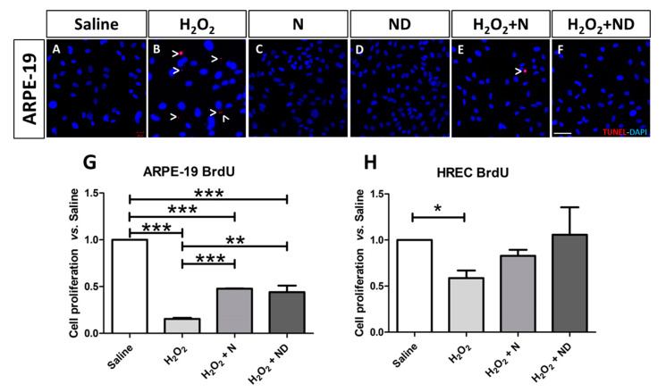
Late apoptosis assessed in ARPE-19 cells by TUNEL labelling and imaged under a confocal microscope. Application of N and ND treatments (62.34 μg/mL) to the media (C,D) showed similar results to saline (A). Addition of H2O2 (2 h; 600 μM) to induce oxidative stress increased TUNEL-positive-stained ARPE-19 cells (B; red), while TUNEL labelling was absent by application of N and ND treatments (62.34 μg/mL each) during the last hour of the induction (E,F). Nuclei were labelled with DAPI (blue). Scale bar: 50 µm. Cell proliferation assay was performed in both ARPE-19 and HREC cells (G,H), n = 3. BrdU expression levels were significantly reduced in both cell lines following oxidative stress-induced conditions by 1000 μM H2O2 for 2h. This effect was significantly recovered in ARPE-19 cells by application of N (p < 0.001) and ND (p < 0.01) treatments (62.34 μg/mL each) during the last hour of the induction, while a strong tendency towards significance was observed in HREC cells (* p < 0.05, ** p < 0.001, *** p < 0.001).

