Fig. 1.
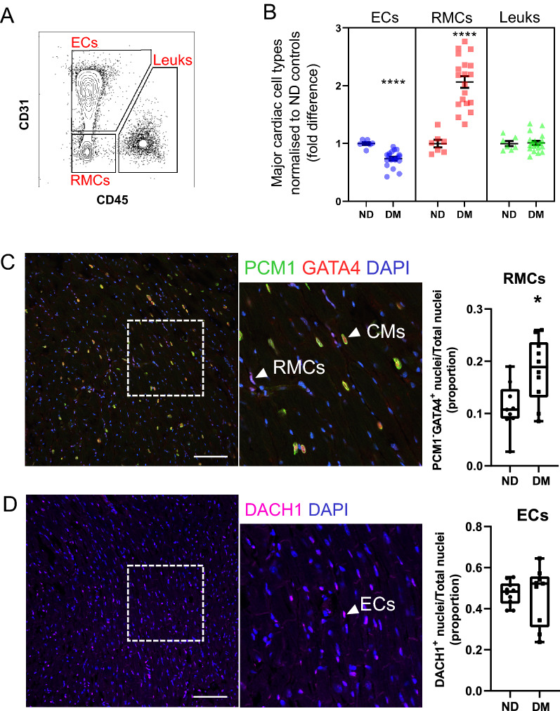
Differences in the abundance of major non-myocyte cell classes in the diabetic heart. A Flow cytometry contour plot displaying gating of major non-myocyte cell types for quantification of cell type proportion (summarised in B). For full gating strategy see Additional file 1: Figure S2. Endothelial cells (ECs; CD31+), resident mesenchymal cells (RMCs; CD31−CD45−) and leukocytes (Leuks; CD45+). B Proportions of major cell types in non-diabetic (ND; n = 7) and diabetic (DM; n = 19) mouse hearts. Individual sample values are shown with mean ± SEM. C Immunohistochemical analysis of the abundance of RMCs in ND and diabetic mouse heart left ventricles. Left and middle panels show representative confocal micrographs of mouse heart tissue stained for PCM1 and GATA4. PCM1+GATA4+ and PCM1−GATA4+ nuclei correspond to nuclei of cardiomyocytes (CM) and RMCs respectively. Nuclei are counterstained with DAPI. Right panel (box-plot) summarises proportion of nuclei corresponding to RMCs in ND (n = 9) vs. DM (n = 10) enumerated from micrographs. Whiskers of box-and-whisker plot indicate max and min. (D) As for C, heart left ventricle sections were stained with DACH1 to identify nuclei corresponding to endothelial cells in ND (n = 10) and DM (n = 9) left ventricles. *P < 0.05, **** P < 0.0001 (Student’s unpaired t-test). Scale bar = 100 µM

