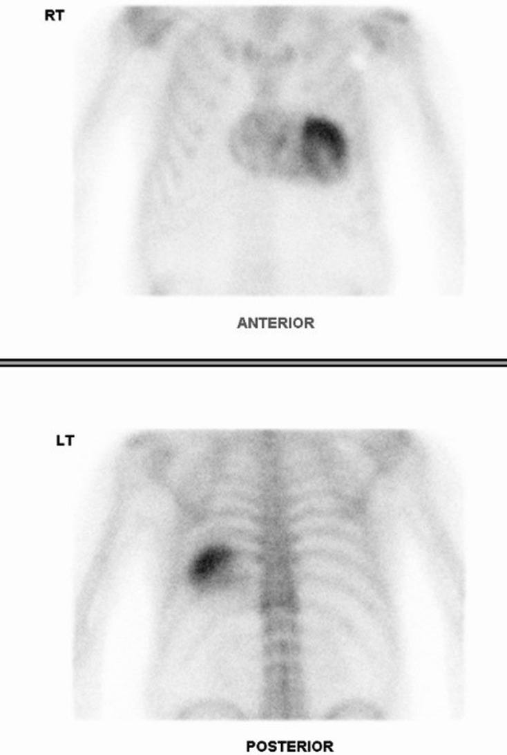
Figure 7.

Cardiac scintigraphy in case 3. Anterior and posterior planar thoracic images, acquired 3 h after 99mTc-DPD administration, demonstrate severe myocardial uptake (Perugini 3 – uptake greater than rib uptake) predominantly in the left ventricle, compatible with the diagnosis of cardiac ATTR amyloidosis. This study was performed 8 years after initial whole-body bone scan and 3/4 years after
