
Figure 1.
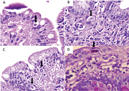
Sections of the duodenum (H&E stain, 1000x). Cystoisospora belli is present inside the epithelium (arrows) with a halo around it (A, B, C). There are also many eosinophils, neutrophils, and lymphocytes in the lamina propria (B). Pink granular staining of parasite with periodic acid-Schiff histochemical staining (D) (PAS, 1000x).