. 2021 Apr 29;12(21):7308–7323. doi: 10.1039/d1sc01886h

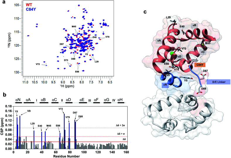
Fig. 2. Mutation-induced human cardiac TnC open-like state. (a) Superposition of 1H–15N HSQC spectra of WT (red) and C84Y (blue) in the primed state. Highlighted residues are those identified as undergoing significant structural changes; (b) chemical shift perturbation (CSP) analysis of C84Y against WT as a function of residue number. Residues deviating from the average values of CSP (Δδ) + 1 S.D. (Δδ + 1σ) and + 2 S.D. (Δδ + 2σ) are highlighted. Secondary content representation (αN–αH) are depicted as horizontal rectangles in the top. Ca2+-binding sites are shown as roman numerals; (c) regulatory (salmon) and structural (gray) cTnC domains show as sticks residues with significant structural changes due to the C84Y substitution. The N-helix (αN) and the D/E linker are colored blue.

Fig. 2