. 2021 Apr 29;12(21):7308–7323. doi: 10.1039/d1sc01886h

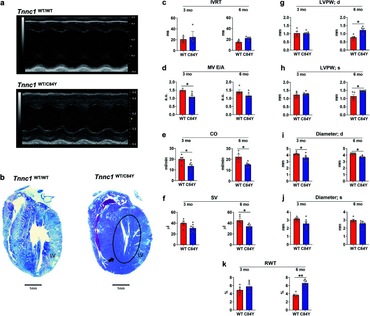
Fig. 5. Knock-in mouse model bearing variant Tnnc1WT/C84Y recapitulates hypertrophic cardiomyopathy. (a) Echocardiography reveals differences in cardiac structure and function in 3 month-old knock-in Tnnc1WT/C84Y mice; (b) representative tissue slices from Tnnc1WT/WT and Tnnc1WT/C84Y mouse hearts stained with Masson's trichrome. Representative, longitudinally sectioned, stained hearts exhibit morphology of advanced cardiac disease in 14 month-old Tnnc1WT/C84Y but not Tnnc1WT/WT mouse hearts. Note the extensive hypertrophy of the left ventricle (LV) in Tnnc1WT/C84Y heart (circled area). (c–k) M-mode and pulsed wave Doppler measurements were analyzed in both the Tnnc1WT/WT and Tnnc1WT/C84Y mice at 3 and 6 months of age. (c)IVRT – isovolumetric relaxation time; (d) MV E/A – mitral valve early peak flow velocity – atrial peak flow velocity ratio; (e) CO – cardiac output; (f) SV – stroke volume; (g) LVPW – left ventricular posterior wall thickness during diastole (d) or; (h) systole (s); (i) diameter during diastole (d) or; (j) systole (s); (k) relative ventricular wall thickness; ANOVA with least significant difference post-hoc test was performed to determine significance. *p < 0.05; **p < 0.001 WT vs. C84Y within age time point. The heart rate was statistically significant lower in Tnnc1WT/C84Y compared to Tnnc1WT/WT (3 months, 504 vs. 450 bpm and 6 months 501 vs. 452 bpm, ESI Table 1).

Fig. 5