
Figure 2.
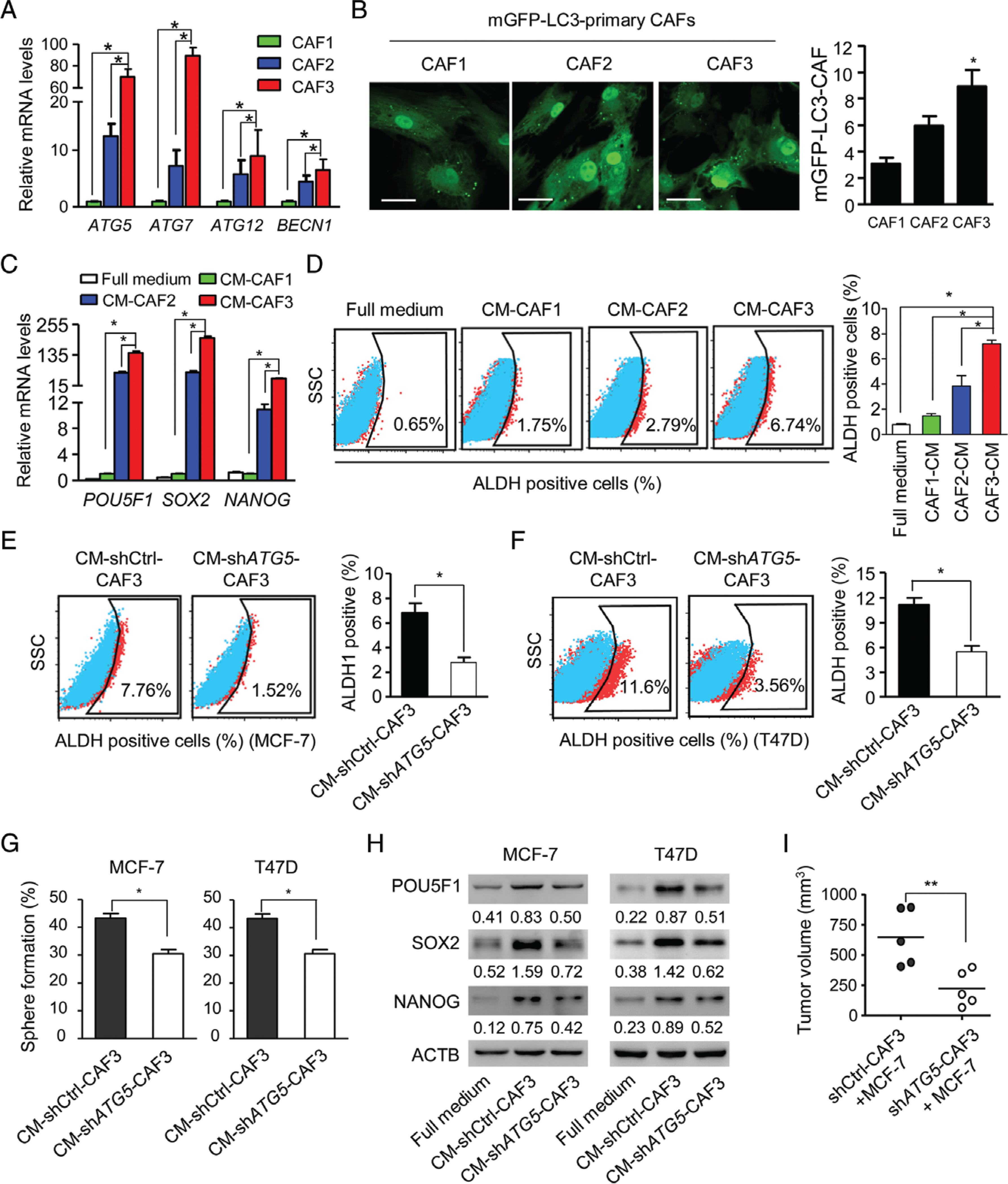
Autophagic CAFs enhance the stemness of luminal breast cancer cells. (A) Quantitative RT-PCR of autophagy gene mRNAs in primary CAFs (CAF1, CAF2, and CAF3) isolated from luminal breast cancers in serum-free medium for 24 h. (B) Primary CAFs transfected with mGFP–LC3 were cultured in serum-free medium for 24 h. Confocal microscopy was used to assess autophagy, as shown by green spots (left panel). Sixty cells were randomly chosen for counting of the number of mGFP–LC3 fluorescent spots per CAF (right panel). Scale bar: 50 μm. (C) The mRNA levels of the stem cell marker genes POU5F, SOX2 and NANOG in MCF-7 luminal breast cancer cells cultured with full medium and supernatants from CAF1, CAF2 and CAF3 in serum-free medium (CM-CAF). (D) ALDH+ cells examined by flow cytometry in MCF-7 cells in full medium and CM-CAFs. (E, F) ALDH activity in MCF-7 and T47D cells cultured in CM from shCtrl-CAF3 (CM-shCtrl-CAF3) and shATG5-CAF3 (CM-shATG5-CAF3). (G) Sphere-forming capabilities of MCF-7 and T47D cells cultured in CM-shCtrl-CAF3 or CM-shATG5-CAF3. (H) Western blots of POU5F1, SOX2 and NANOG in MCF-7 and T47D cells cultured in full medium, CM-shCtrl-CAF3, and CM-shATG5-CAF3. (I) Co-injection of MCF-7 cells with shCtrl-CAF3 or shATG5-CAF3 into the mammary pads of NOD-SCID mice. Tumours were collected 4 weeks after implantation. Dot plots show tumour volumes (n = 5 mice in each group). *P < 0.05; **P < 0.01.